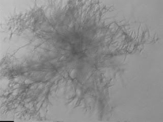
Fungal contamination is normally the easiest to see, as in advanced cases it can be seen with the naked eye. They normally look kinda whitish/yellowish and fluffy.

Bacterial contamination is a little harder, but can be seen under a high powered microscope as little black dots that move around the cells. Not a good sign. They also cause a sudden change to the pH of cell growth medium (and therefore the colour!) from redish/pink to yellow
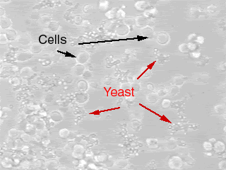
Yeast contamination is also visible under high powered microscopy. Must admit I've never seen yeast contamination myself, however they look like small ovals that bud of from each other.


The hardest contamination to detect is mycoplasma, mainly because it cannot be detected by light microscopy. In fact, we dont even test for this in the lab! Thought it normally has to come from an infected cell source (which we dont have, so I doubt this will affect us!)
Argh! I am trying deperately to save my cells, I'm hoping tomorrow they'll be looking better... But i doubt it. And if they die I'll have to thaw out a new crop!
Darn I hate cell culture!
Argh! I am trying deperately to save my cells, I'm hoping tomorrow they'll be looking better... But i doubt it. And if they die I'll have to thaw out a new crop!
Darn I hate cell culture!


1 comment:
I know this is an old post but the same thing is happening to my HEK cells. did you ever find out what it was?
Post a Comment